The higher vascular plants possess a large variation in the arrangements and structure of the primary tissues in their roots, stems and leaves.
Primary tissues in the dicotyledonous stem
Anatomically, the stem possesses a dermal tissue system from where the epidermis, vascular tissue system develops. Next vascular bundles and the ground tissue system originate from this vascular tissue system and rest of the tissues originate from the ground tissue system which is found in the stem. All these primary tissues show a great diversity in their structure and arrangements.
Epidermis:
The epidermis is the peripheral protective cell layer in the stem. The compactly arranged cells of this layer are more or less tubular or rectangular with these primary walls. The outer cell surface of this layer is often covered by a layer of cuticle or sometimes contains chlorenchyma cells to perform the function of photosynthesis.
Cortex:
This tissue system is situated in between the epidermis and the stele. Thin-walled parenchyma cells formed this layer, e.g., salicornia, Pelargonium, etc.
Endodermis:
It is the inner delimiting layer of the cortex. This layer is inconspicuously demarcated in aerial stems with the exception in Piper where Casparian strips are also observed. Endodermal cells deposit lignin and suberin in addition to cellulose on their transverse and radial walls as stripes or bands to form a Casparian strip or band.
Functions of endodermis –
- Fat, starch, protein, and tannins are accumulated in these cells.
- Casperian strips restrict the movement of water, iron, lead and copper in either direction.
- These cells form a barrier for pathogens as it contains a large number of quinones that inhibit fungal and bacterial growth.
- Sometimes the cork cambium or phellogen originates from endodermis ( stem of Cocculus, Paederia foetida).
Stele:
The stele is the central core portion of the vascular and ground tissue systems where endodermis encircled this portion. Ground tissue system is formed from the parenchymatous pith. The pith cells either lignified or pitted form.
Primary tissues in the monocotyledonous stem
Monocotyledonous stems also contain the dermal, vascular and ground tissue systems. Dermal tissue system forms epidermis, the vascular tissue system consists of vascular bundles and the ground tissue system consists mainly of parenchyma.
Epidermis:
In this type of epidermis, it is usually devoid of glandular and non-glandular trichomes.
Ground tissue:
In monocot stems, there is not found any distinction between cortex and pith in the ground tissue. Distinction found sometimes in Asparagus and Amaryllidaceae families.
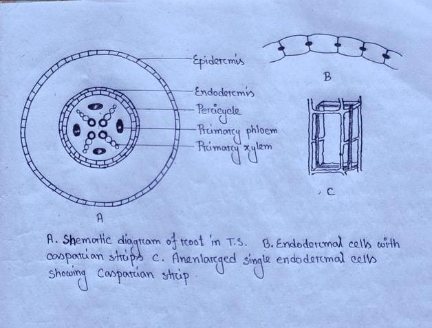
Arrangements of primary tissues in roots
A cross-section of root reveals the following tissues:
Epiblema:
Epiblema is the dermal tissue system, also called rhizodermis. Elongated, thin-walled cells constructed the layer. A thin cuticle present on the cell wall, devoid of intercellular spaces. The most important feature is the presence of unicellular and unbranched root hairs that are projections of certain cells of this layer. The function of root hairs is anchorage means it absorbs water and mineral salts from the soil solution. The root hair is formed from some special epidermal cells with smaller size and dense cytoplasm. These specialised cells are called trichoblasts or piliferous cells. Thus it is termed a piliferous layer.
Epiblema typically single-layered with the exception in the aerial roots of certain epiphytes where it is multilayered. This multiple epidermises termed as velamen, contents several layers of dead cells externally forming a sheath around the root. It protects thin-walled cortical cells from dedication. The innermost layer of the velamen is known as exodermis, considered as the outermost layer of the cortex.
Cortex:
This portion is mainly parenchymatous, where schizogenous and lysigenous intercellular spaces may be present. In case of hydrophytes, also seen aerenchyma when root cortical cells are very regularly arranged. The cortex portion of roots is usually very wide and the cells often contain starch, crystals etc. Collenchyma rarely occurs in the root cortex ( Monstera) and the cortical cells of many epiphytes contain chloroplasts.
Endodermis:
The innermost layer of the cortex is the endodermis that encircles the stele. In roots devoid of secondary growth suberin deposits as lamellae over the whole inner primary wall of the endodermal cells including Casparian strips and afterwards cellulose is deposited centripetally on the inside of suberin lamellae ( Iris). Thus the endodermal cells are sometimes thickened around their radial walls. In the protoxylem adjacent, endodermal cells thickening is either delayed or there is no thickening except the Casparian strips. These cells are known as passage cells, through which water and mineral matters get entry into the xylem from cortical cells.

Stele:
Stele consists of radial vascular bundles and pith. Just beneath the endodermis and external to the vascular tissues, there is a cell layer called pericycle. This layer is mostly uniseriate but sometimes multiseriate (Agave, Smilax, Salix). Beneath the pericycle alternatively arranged the primary xylem and phloem. The primary xylem contains thick-walled, lignified tracheary elements that mature centripetally. Here, xylem type is exarch means protoxylem lies on the peripheral side and metaxylem lies towards the center portion. Centripetal differentiation is also observed in the phloem.
Arrangements of tissues in leaves
Leaves also consist of three main tissue systems – the dermal tissue system, the ground tissue system and the vascular tissue system. The dermal tissue system consists of the upper and lower epidermis, the ground tissue system comprises the mesophyll and the vascular tissue system is represented by the vascular bundles.
Epidermis:
This is the uniseriate outermost layer. In the case of Ficus, Piper, Nerium multiseriate epidermis shows xerophytic characteristics and protects inner tissues. Beside this, where both the epidermis are multiseriate the upper one consists of more layers than the lower. In multiple lower epidermis is a sub-stomatal cavity present beneath the stoma (Piper). In Nerium the lower epidermis is multiseriate. The stomata are present in stomatal chambers only where the epidermis is uniseriate. This type of stomata is called sunken stomata. Stomata may occur either on the upper epidermis ( Nymphaea) or on the lower epidermis ( Ficus) or both sides.
Mesophyll:
It is the parenchyma ground tissue of leaf lying internal to the epidermis. In dorsiventral leaves, these parenchymas are of two types: palisade parenchyma and spongy parenchyma.
In leaf t.s. The palisade cells appear as elongated cylindrical structures and in l.s. They appear as spherical structures. Palisade parenchyma is present on both sides but spongy parenchyma remains in between in the form strips. If the palisade parenchyma occurs on the dorsal surface only the leaf termed as dorsiventral whereas a leaf is said to be isobilateral when the spongy parenchyma occurs on both surfaces.
Vascular bundles:
This is situated at the centre with the inverse orientation of vascular tissues. Xylem occurs on the adaxial side and phloem on the abaxial side. Vascular bundles are sheathed by parenchyma cells morphologically distinguishable from the adjacent mesophyll cells ( in Cyprus, Saccharum, Zea etc). These parenchyma cells are usually larger, thick-walled and contain chloroplast, this parenchyma sheath is termed as bundle sheath.
If you have any query regarding “What are the Primary Tissues?”post, then don’t hesitate to coment below.

